SIXPAD의
효과 검증
EMS의 올바른 이론・효과를
세상에 널리 올바르게 알리기 위한 노력
SIXPAD의 생체 효과 검증
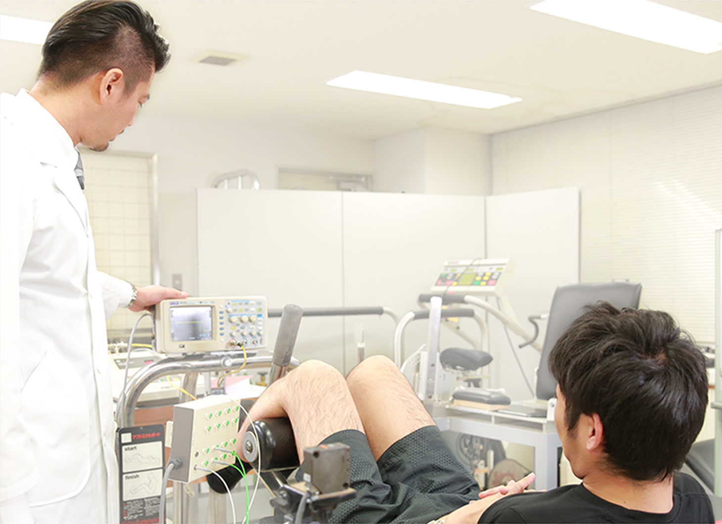
주쿄대학 국제교양학부 와타나베 코헤이 부교수와 함께 SIXPAD 트레이닝(근전기 자극)으로 근육 수축이 이루어지고, 생리학적으로 근피로가 일어날 수 있는지를 검증하였습니다.

학회 발표
2015년 7월 5일 '제16회 일본전기생리운동학회(JSEK)'에서 MTG는 교토대학 모리타니 토시오 명예교수, 주쿄대학 국제교양학부 와타나베 코헤이 부교수와 함께 전기 자극 장치에 의한 근피로 특성 검증의 성과에 대하여 학회 발표를 실시하였습니다.
Proceedings of the 16th Japan Society of Electrophysiology and Kinesiology (JSEK)
Neuromuscular fatigue induced by
wearable small size electrical stimulation device
Shuhei Kawade1, Kohei Watanabe2, Toshio Moritani3
1MTG Co., Ltd., 2School of International Liberal Studies, Chukyo University,
3Human and Environmental Studies, Kyoto Universityy
1 Introduction
Electrical muscle stimulation (EMS) has been used in clinical settings for a long time in fields like nursing care and rehabilitation as an alternate means of exercise for people who cannot move of their own volition.
In recent years, electrical muscle stimulation has come to be widely used by even healthy individuals to improve their lack of exercise or to maintain and/or improve their health. We anticipate that in the future it will be used both in everyday life and for voluntary exercise. Further, a significant training effect (increase in maximum muscle strength) has been reported by adding EMS to normal resistance training and jump training. [1] In addition, a new form of exercise has been proposed in which EMS would induce a specific metabolic response by incorporating EMS into aerobic exercise. [2]
However, most of the EMS devices on the market have a lot of cables between the EMS device and the person exercising; this is because the unit itself, the controller and electrodes are separate, as well as due to the need for a power supply. If the unit and the controller are large, it is difficult to use them during activities in everyday life or during different exercises, so we anticipate the development of EMS devices that miniaturize and integrate the unit, controller and electrodes.
And because it is important to make the device wearable, such as by using batteries for the power supply, in order to eliminate restrictions on the person exercising, a concern is that this will decrease the muscle output induced by the lower strength of the stimulation.
2 Purpose
The aim is to evaluate the characteristics of muscle fatigue associated with muscle contractions induced by an EMS device that has been miniaturized and made wearable.
3 Method
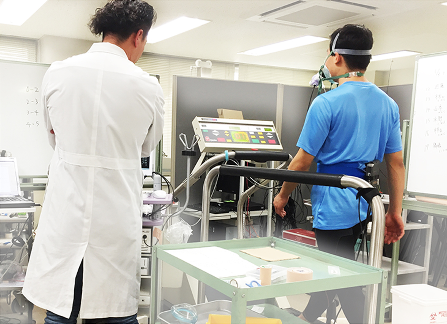
The research has the approval of an “Ethics Review on Research using Human Subjects” at Chukyo University (Application No. 2014-001) Further the content of measurements was explained to subjects ahead of time, their informed consent obtained in writing and the experiment conducted.
The subjects were 12 males (ages: 21.4 ±1 year; height: 173.0 cm ±7.0 cm; Weight: 62.0 ± 5.8 kg; BMI: 20.7 ± 1.5).
The test site was the bicep, which was measured to evaluate the muscle fatigue due to electrical stimulation and voluntary movement.
- (1) Measurement of maximum muscle strength (MVC-PRE)
- The right arm of the subject was measured with the arm fixed at a joint angle of 120 degrees (interior angle) using an isometric elbow joint flexion strength measurement device (made by VINE).
- (2) Measurement of surface EMG median frequency (MF-PRE)
- Using an electromyograph (EM-272, made by Noraxon), a surface EMG was taken with twin leads proximal to the bicep muscle while it was exhibiting submaximal muscle strength (maintaining 50% of MVC for 3 seconds) and was recorded (AB-611J, made by Nihon Kohden).
- (3) Exercise load
- “Electrical stimulation trial”
An electrical skeletal-muscle stimulation device (made by MTG) was attached proximally to the bicep muscle, which was stimulated electrically for approximately 20 minutes while held with an elastic band. The maximum stimulation subjects could withstand was specified, which was the maximum stimulation strength of the device (40V). The stimulation frequency was at 20 Hz, which was applied continuously with 3 seconds of contraction interspersed with 2 seconds of rest.
“Voluntary movement trial”
Voluntary movement at the same level as that induced by electrical stimulation was specified, so voluntary movement (isometric elbow joint flexion movement) that exhibited the muscle strength of about 12% of maximum muscle strength was conducted for approximately 20 minutes, with 3 seconds of contraction interspersed with 2 seconds of rest. - (4) Measurement (MVC-POST) of maximum muscle strength after the application of load to the muscle
- This was made in the same way as (1).
- (5) Measurement (MF-POST) of surface EMG median frequency after the application of load to the muscle
- This was made in the same way as (2).
- Based on the results of the foregoing, the rates of change of maximum muscle strength (MVC-POST/MVC-PRE) and that of EMG median frequency (MF-POST/MF-PRE) before and after application of load to the muscle were calculated.
4 Results
The rates of change of maximum muscle strength (MVC-POST/MVC-PRE) were 71.9% ± 5% for the electrical stimulation trial and 93.2% ± 4.2% under the voluntary movement trial, so maximum muscle strength under the electrical stimulation trial dropped significantly more (p <0.05).
The rates of change (MF-POST/MF-PRE) of EMG median frequency during exhibition of submaximal muscle strength were 87.0±15.7% for the electrical stimulation trial and 105.5±9.2% under the voluntary movement trial, so it dropped significantly more under the electrical stimulation trial (p <0.05).
5 Discussion
Because a drop in maximum muscle strength is an indication of muscle fatigue, despite the muscle output being similar, the results we obtained suggest that muscle fatigue is accelerated more by electrical stimulation than by voluntary movement.
The drop in EMG median frequency is well-known to reflect fatigue, especially peripherally (muscle itself). A drop in the median frequency was not observed with voluntary movement in this study. This fact points to the possibility that almost no peripheral fatigue was induced by voluntary movement during the 20 minutes provided.
By contrast, a drop in the median frequency was observed under electrical stimulation, despite it being such low-intensity exercise. From an electrophysiological standpoint, this supports the possibility that electrical stimulation gives rise to greater fatigue than voluntary movement suggested from the drop in maximum muscle strength. We can also confirm this from the fact that almost no fatigue is produced when voluntary movement is at the level of 10% of maximum muscle strength. Despite such an extremely low level of intensity in muscle output, we obtained a result that reflects that electrical stimulation gives rise to fatigue. It is thought that this is due to different motor units and/or muscle fibres being mobilised during voluntary movement versus electrical stimulation.
According to the principle of size, under voluntary movement, muscle fibres are mobilised in order, starting from the low-tension muscle fibres (so-called slow-twitch muscle) that are not readily fatigued by low-intensity exercise. On the other hand, high-tension muscle fibres (so-called fast-twitch muscle) that do fatigue easily are basically only mobilised during high-intensity exercise. It is known that electrical stimulation of skeletal muscles, the motor neurons that control them, or both, mobilises motor units randomly, which does not follow this principle of size. [3] [4]
In other words, fast-twitch muscles that are not mobilised under low-intensity voluntary movement are mobilized under electrical stimulation, even if the muscle output is low-intensity. As was observed during this study, even though under low-intensity voluntary movement is mobilised, fatigue is not produced, the low intensity movement is what caused an increase in fatigue under electrical stimulation. From the foregoing results, the inference is that the wearable EMS device used in the study can induce specific muscular fatigue via EMS.
References
- [1] Herrero et al. Journal of Strength and Conditioning Research volume24:1609-1615, 2010
- [2] Watanabe et al. Eur J Appl Physiol 114:1801-1807,2014
- [3] Gregory CM, Bickel CS. Phys Ther 85:358-364,2005
- [4] Maffiuletti et al. Eur J Appl Physiol 111:2391-2397,2011
Training Suit의 효과 검증
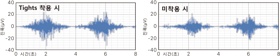
근육 활동 레벨의 증가를 입증.
검증 항목
Tights 착용 시와 미착용 시에 같은 동작(무릎 관절을 180도에서 90도로 구부리는 과제)을 수행하여 햄스트링의 활동 전위를 비교하였고, 그 결과 착용 시에는 근전도 진폭값이 약 30%* 증가하여 미착용 시에 비해 더 많은 신경근 활동이 이루어진다는 것을 확인할 수 있었습니다. 이는 같은 운동이라도 햄스트링이 더 강한 운동을 수행하려고 한다는 것을 반영한다고 해석할 수 있습니다.
*피험자 10명(31세~50세 남성)의 Tights 착용 시와 미착용 시의 근전위 진폭값의 평균값 비교(제3자 기관 조사)
햄스트링 표면 근전도 파형의 일례
실험 데이터 해석 협력 :와타나베 코헤이 주쿄대학 국제교양학부 준교수

주쿄대학 국제교양학부와타나베 코헤이 준교수
- 2010년나고야( 名古屋 )대학대학원 교육발달과학연구과 박사 후기과정 수료
- 2010년교토대학대학원 인간・환경학연구과 특별연구원
- 2012년주쿄대학 국제교양학부 준교수
일본체육학회, 일본체력의학회, 일본바이오메카닉스학회, 일본운동생리학회, 일본전기생리운동학회, 미국스포츠의학회(ACSM), 국제전기생리운동학회 소속. 전문분야는 운동생리학・생체역학이며, 운동 중의 신체의 각각의 기능변화와 적응에서 ‘골격근과 이를 지배하는 운동신경’에 초점을 맞춰 인간을 대상으로 연구.